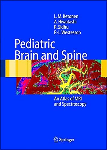
By Per-Lennart Westesson, Leena M. Ketonen, Akio Hiwatashi, Ravinder Sidhu
This sensible MR imaging atlas covers all features of pediatric mind and backbone imaging together with modern thoughts resembling diffusion imaging, fetal imaging and MR spectroscopy. The publication contains 12 chapters, beginning with the constructing pediatric mind and carrying on with with metabolic, congenital, genetic, stressful, inflammatory and neoplastic methods of the mind and backbone. every one entity is comprehensively illustrated with a number of most fulfilling photos and followed by means of medical heritage, a quick dialogue and the main pertinent references. There are wide illustrations of MR imaging of kid abuse. The authors are at present all on the collage of Rochester, big apple, yet have lengthy event of pediatric neuroradiology from a world point of view, which promises a large view of the topic. The publication is efficacious for radiologists, pediatricians, neurologists, neurosurgeons, neuroscientists and pediatric neurologists, in addition to for physicians in education in those parts. This richly illustrated atlas is a short medical reference for all excited by pediatric neuroimaging.
Read Online or Download Pediatric Brain and Spine: An Atlas of MRI and Spectroscopy PDF
Similar pediatrics books
Understanding Developmental Language Disorders: From Theory to Practice
Developmental language issues (DLD) ensue while a baby fails to improve his or her local language frequently for no obvious cause. not on time improvement of speech and/or language is among the most typical purposes for folks of preschool little ones to hunt the recommendation in their family members general practitioner. even though a few young ones swiftly increase, others have extra continual language problems.
Toward an Integrated Science of Research on Families: Workshop Report
Demographic alterations, immigration, monetary upheavals, and altering societal mores are growing new and adjusted constructions, approaches, and relationships in American households this present day. As households suffer swift swap, kinfolk technological know-how is on the breaking point of a brand new and intriguing integration throughout tools, disciplines, and epistemological views.
Pediatric Infectious Diseases for the Practitioner
Accomplished Manuals in Pediatrics are designed to expand the prac titioner's medical scope through supplying a variety of diagnostic and administration talents usually thought of to be the particular area of the experts. even supposing the sequence as an entire constitutes a complete textual content in pediatrics, every one quantity stands by itself as a self-contained reference for the busy practitioner.
Practitioner’s Guide to Behavioral Problems in Children
Over the past 25 years of scientific perform, i've been inspired with a paradox, specifically, the distinctiveness in every one baby, not like the common commonalities present in the improvement of behavioral difficulties. i've got additionally been duly inspired with the resilience of youngsters and their households, and the impression that provision of data relating to improvement and behaviour may have on facilitating this resilience.
- Understanding Asperger Syndrome and High Functioning Autism (The Autism Spectrum Disorders Library)
- Clinical child neuropsychiatry
- The Complexity of Adolescent Obesity: Causes, Correlates, and Consequence
- Pediatric nursing care plans for the hospitalized child
- All in Your Head: Making Sense of Pediatric Pain
Additional info for Pediatric Brain and Spine: An Atlas of MRI and Spectroscopy
Example text
The lateral ventricles are widely apart and the frontal horns are pointed 20 Chapter 2 C. T1-weighted image shows agenesis of corpus callosum and the midline cyst. The fontal horns are wide apart (arrowheads) because if the high-riding third ventricle. Subependymal nodular heterotopia is also noted. Note the pointed forehead in this patient with metopic suture closure seen on CT D. Coronal SPGR image reveals the typical “Viking helmet” or “moose head” configuration of the ventricles and high-riding third ventricle.
Heterogeneous enhancement is seen in the medial temporal lobe B. Sagittal T1-weighted image reveals dumbbell shaped brain tissue (arrow) herniating inferiorly from the cranial cavity to the oropharynx C. Coronal T1-weighted image shows the herniating brain tissue in the oropharynx (arrow), left to the clivus. The signal is heterogeneous corresponding to the hamartomatous changes seen in pathology. A large CSF space is seen lateral to this abnormal brain tissue within the cranium. A normal hippocampus is not identified in this side A 5-month-old female with neck and parapharyngeal mass that during later surgery proved to be brain tissue with hamartomatous areas.
Schizencephaly may be inherited or may be a result of an early intrauterine insult, such as exposure to toxins, infection or trauma. Suggested Reading Barkovich AJ, Norman D (1988) MR imaging of schizencephaly. AJR Am J Roentgenol 150:1391–1396 Packard AM, Miller VS, Delgado MR (1997) Schizencephaly: correlations of clinical and radiological features. 18 Lissencephaly A B Discussion Lissencephaly Clinical Presentation X-linked lissencephaly. A 3-year-old male with microcephaly, profound developmental delay and seizures.